
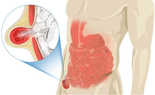
A hernia is a condition where an organ, often the intestine, protrudes out of the abdominal cavity through a fragile area of the abdominal wall. There are several sites where the intestine can protrude, such as the groin area (Groin Hernia) or the abdominal wall (Abdominal Hernia). This condition is a health issue that can significantly interfere with daily life and should not be overlooked.
The protrusion of an organ appears as a bulging lump. If the condition is not severe, the patient can often push the contents of the hernia sac back into the abdominal cavity themselves (reducible). Pain may occur when the lump protrudes. This pain is caused by the edge of the hernia sac in the abdominal wall constricting the contents, especially the intestine and omentum (fatty tissue).
The pain typically subsides when the organ in the hernia sac is pushed back into the abdominal cavity. In cases where the protrusion occurs and the contents cannot be pushed back (incarcerated hernia), there will be severe pain in the hernia area because the organ is constricted by the edge of the hernia sac. Specifically, the intestine can swell and lose blood supply. When this happens, emergency surgery is required to prevent intestinal tissue death due to lack of blood (strangulated hernia).
This is a condition where a portion of the internal organs—most commonly the intestinal wall (especially the small intestine) and theomentum (a double layer of fatty tissue that covers the abdominal organs, serving as a structural component that holds various organs together in the abdominal cavity)—moves or pushes out of its original position through a hole or a weakened abdominal wall (consisting of the muscle layer and fascia sheet). The protruding contents are then covered by the hernia sac, which includes a thin muscle layer, fat, and skin.
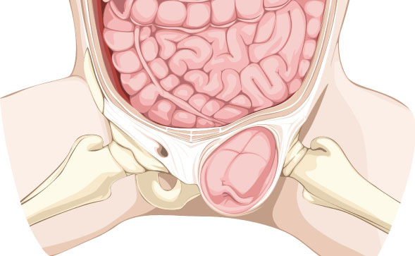
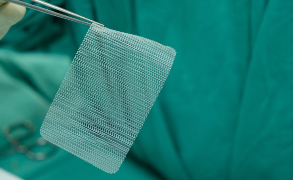
This is the most common type, accounting for about 75% of all hernias. In men, a lump is often felt in the groin or scrotum area. It is caused by weakness in the abdominal wall near the groin. Laparoscopic surgery involves small incisions, less pain, and fast recovery, offering an effective alternative treatment option. Laparoscopic Inguinal Herniorrhaphy is one such successful surgical procedure. It uses a technique where a mesh is placed to cover the hernia defect from inside the abdominal wall, inserted through small incisions (typically below the navel). A synthetic three-dimensional mesh is used to strengthen and close the hernia defect.
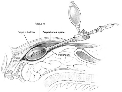
This procedure is performed through the space behind the abdominal muscle layer and the peritoneum (the lining of the abdominal cavity). It is the most popular laparoscopic method because it avoids entering the abdominal cavity, leading to fewer complications. This type of surgery yields good results and has a low recurrence rate. However, the laparoscopic surgeon needs a significant learning curve, requiring experience with 50–100 cases or more, to achieve optimal surgical outcomes.
This method involves pulling the hernia sac and its contents back into the abdominal cavity and then placing a mesh outside the peritoneum. The procedure typically involves three small incisions: a 1 cm incision below the navel for the scope and 0.5 cm incisions for instruments. During the surgery, the surgeon expands the space between the back of the abdominal wall and the peritoneum to clearly visualize the hernia defect from behind the abdominal wall—an advantage over conventional open hernia repair. A synthetic mesh, which is currently developed to fit the inner abdominal anatomy (anatomical 3D mesh), is then placed to cover the weakened muscle and fascia. The mesh is fixed using tacks, which come in various options such as metal, dissolvable materials, and the latest innovation, fibrin glue (which is effective and minimizes post-operative pain more than other materials).
Comparing laparoscopic surgery with traditional open abdominal surgery, laparoscopy for various types of hernias, particularly recurrent cases of inguinal hernia or bilateral groin hernias, offers better outcomes than the open method. It helps patients recover quickly, with minimal wound pain or bruising after the operation.
The hospital stay is typically only 2–3 days. This shorter recovery period allows patients to return to work or normal life sooner than before.